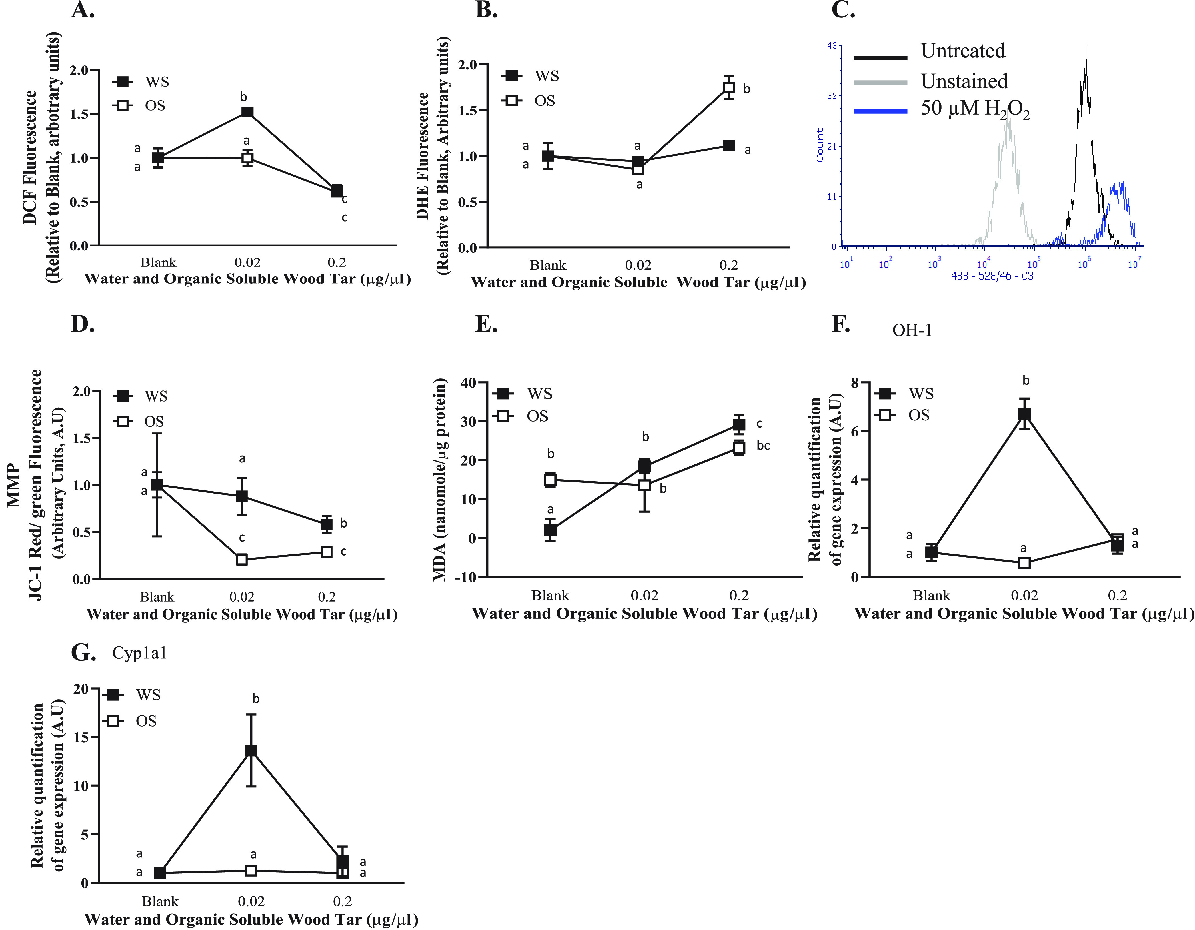
Figure 4.

Wood tar extracts induced oxidative stress alterations in A549 lung epithelial cells. Lung epithelial cells were exposed to water-soluble (WS) or organic-soluble (OS) wood tar extracts at concentrations of 0.02, 0.2, or 1 mg/mL for 5 h. (A) Intracellular ROS were measured using H2DCF-DA, detection was performed by flow cytometry, and 100 μM hydrogen peroxide was used as positive control. (B) Superoxide anions were measured using DHE, detection was performed by flow cytometry, and 100 μM antimycin A was used as positive control. (C) Flow cytometry histogram indicating unstained, untreated, and 100 μM hydrogen peroxide as controls. (D) MMP was measured using JC-1 probe, detection was performed by flow cytometry, and FCCP was used as positive control. (E) Lipid peroxidation was measured in cells homogenates and was calibrated to protein levels examined by Bradford protein assay. Transcription levels were analyzed by real-time PCR for (F) HO-1 and (G) Cyp1a1. β-Actin and HPRT were used as endogenous controls. The data represent the mean ± SD. Means with different letters are significantly different at p < 0.05 using the Tukey HSD test. These experiments were performed in triplicate and were repeated twice.
