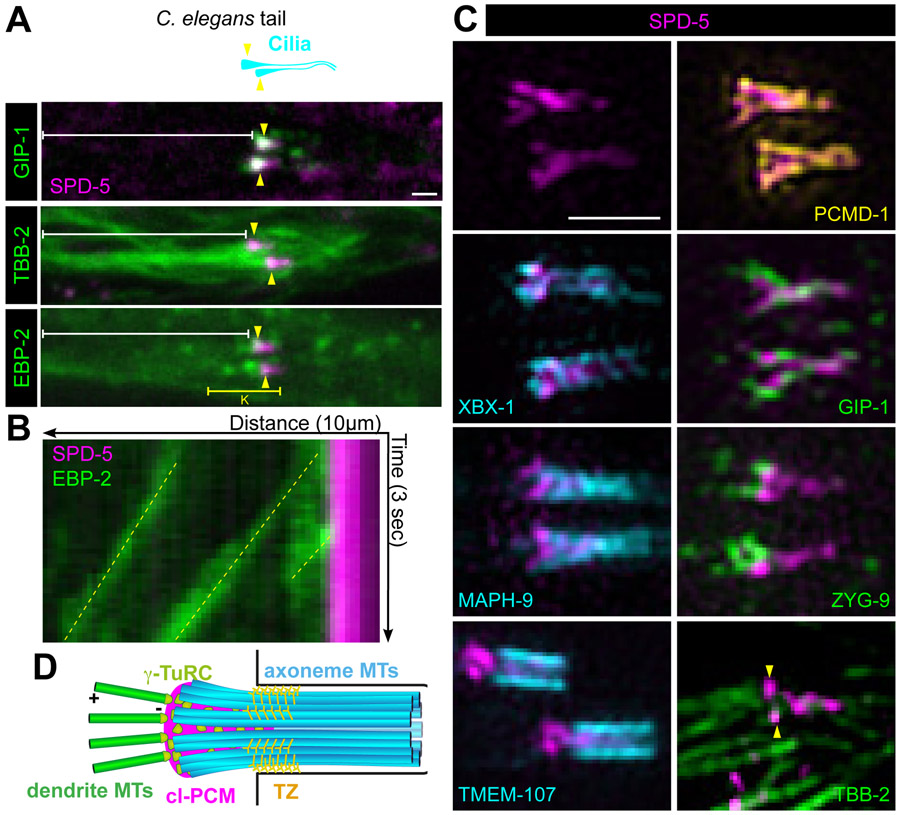
Figure 3. Centriole-less PCM at the ciliary base serves as a MTOC.
(A) Cartoon (top) and images from live adult worms (bottom) of the C. elegans phasmid cilia (cyan) at the tip of the dendrite (white bracket) with ciliary base indicated (yellow arrowhead). (B) Kymograph of yellow bracketed region in A of EBP-2 comets (green, yellow dotted line) traveling from the ciliary base (SPD-5, magenta) toward the cell body. (C) 3D-SIM analysis of SPD-5 (magenta) localization at the ciliary base in phasmids cilia relative to PCMD-1 (yellow), axonemal (cyan), and microtubule-related (green) proteins; Yellow arrowheads indicate centriole-less PCM and white arrowheads indicate cytoplasmic microtubules. (D) Cartoon summarizing the organization of the ciliary base as revealed by 3D-SIM. Axonemal MTs (axMTs, cyan), transition zone (TZ), centriole-less PCM (cl-PCM, magenta), and dendrite microtubules (dendrite MTs, green) are indicated. Scale bars = 1μm.
See also Figure S2.

