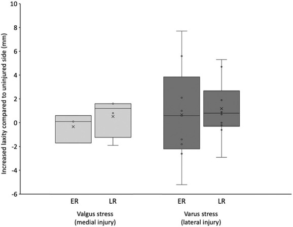
Fig. 3.

This graph shows an assessment of coronal stability using varus and valgus stress radiographs. Lateral-sided injuries were assessed with varus stress, and medial-sided injuries were assessed with valgus stress. The total distance between the relevant articular surfaces was measured and compared with that of the contralateral, uninjured leg. Radiographs were taken at the 1-year follow-up visit by the primary surgeon. Images were evaluated by a blinded assessor; ER = early rehabilitation; LR = late rehabilitation.
