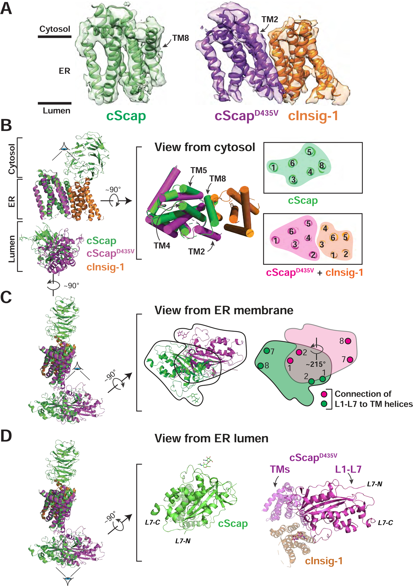
Figure 6. Conformational Differences between cScap and cInsig-1-bound cScapD435V.

(A) Transmembrane domain models and masked cryo-EM density for the cScap and cScapD435V/cInsig-1 structures. cScap is colored green, cScapD435V is colored purple, and cInsig-1 is colored orange.
(B) Left: cScap and cScapD435V-cInsig-1 superimposed based on the TM helices of cScap. The Scap CTD (resolved only in the cScap structure) and the L1-L7 domains are dimmed to highlight the superposition of the transmembrane domains. cScap is colored green, cScapD435V is colored magenta, and cInsig-1 is colored orange. Middle: The superimposed TM helices, rotated 90° with respect to the view shown at left, as viewed from the cytosol. TM helices are shown as cylinders. Right: Slice of the transmembrane domains from the approximate center of the membrane. Numbers indicate the assigned TM helix. Left and middle panels were generated using Pymol, right panel was generated using UCSF Chimera X.
(C) Left: cScap and cScapD435V/cInsig-1 superimposed based on the TM helices of Scap. The Scap CTD (resolved only in the cScap structure) and the TM helices are dimmed to highlight the different L1-L7 position. cScap is colored green, cScapD435V is colored magenta, and cInsig-1 is colored orange. Middle: The superimposed L1-L7 domains, rotated 90° with respect to the view at left, as viewed from the ER membrane. Black trace outlines the outer boundaries of the L1-L7 domain for each structure. Right: The outlined regions from the Middle panel are colored green or magenta corresponding to the WT and D435V (Insig-bound) forms of cScap, respectively. The grey region denotes the overlap of L1-L7 domains. These two domains are related by a rigid body rotation of ~215° about the membrane normal. Approximate positions where L1 and L7 attach to TM helices (TM1 and TM2 for L1; TM7 and TM8 for L7) are indicated by circles.
(D) Left: Superposition of cScap and cScapD435V/cInsig-1 as in (C). Middle: cScap, rotated 90° with respect to the view at left, as viewed from the ER lumen. cScap is colored green. TM helices extending back from the page are dimmed. Right: cScapD435V/cInsig-1 complex, rotated 90° with respect to the view at left, viewed from the ER lumen. cScapD435V is colored magenta and cInsig-1 is colored orange. All TM helices extending back from the page are dimmed.
