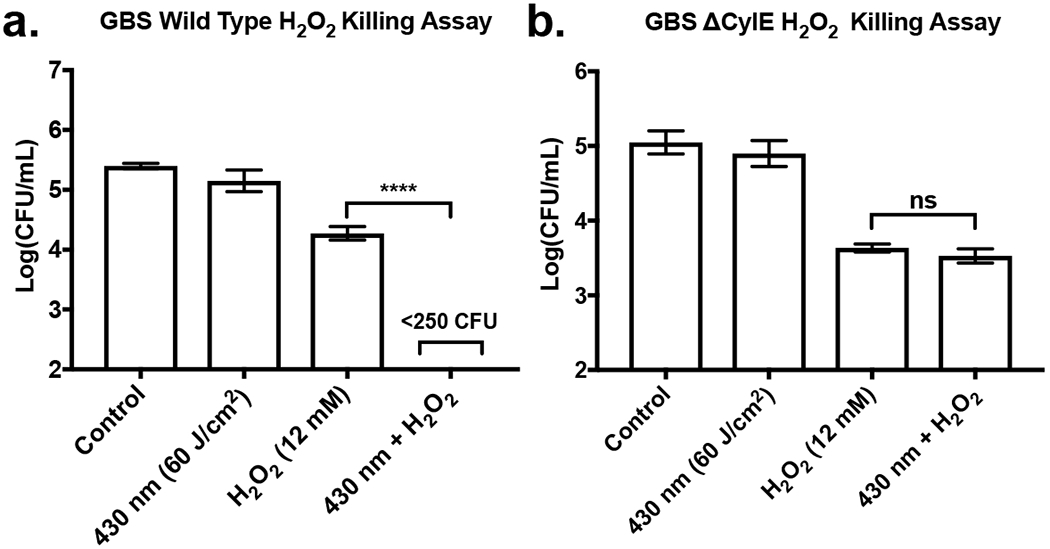
Figure 3.

H2O2 killing assays of wild type and mutant GBS, exhibited through differences in mean bacterial CFU population and standard deviation following incubation within H2O2 environment. (a) Pigment expressing GBS exposed to 60 J/cm2 of 430 nm nanosecond pulsed light and incubated with 12 mM of H2O2 for 1 hour. Combination of 430 nm light exposure and H2O2 resulted in eradication of GBS. (b) Pigment deficient ΔCylE GBS exposed to 60 J/cm2 of pulsed light and incubated with 12 mM of H2O2 for 1 hour. Combination of 430 nm light exposure and H2O2 resulted in no significant improvement in H2O2 antimicrobial activity. ****: p < 0.0001.
