
Figure 4.
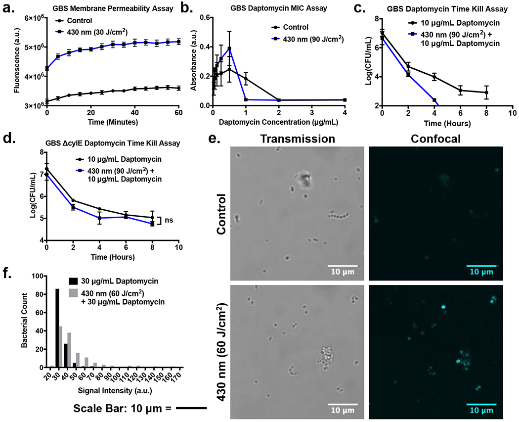
Granadaene photobleaching facilitates cellular uptake and antimicrobial activity of daptomycin. (a) Membrane permeability assay of GBS exposed to 430 nm nanosecond pulsed light utilizing SYTOX Green. Instantaneous increase in SYTOX Green fluorescence following light exposure indicates immediate membrane permeabilization. (b) Minimum Inhibitory Concentration (MIC) of daptomycin on GBS photobleached with blue light decreases 2-fold from 2 μg/mL to 1 μg/mL. (c) CFU count of time killing assay of GBS exposed to 90 J/cm2 of pulsed blue light. GBS exhibits faster response to daptomycin following initial light treatment. (d) CFU count of time killing assay of pigment deficient ΔCylE GBS exposed to 90 J/cm2 of pulsed blue light and treated with 10 μg/mL for 8 hours. Light exposed ΔCylE GBS lacks the same daptomycin enhancement observed in the pigmented strain. (e) Confocal imaging of light exposed GBS treated with 30 μg/mL of daptoymcin-BODIPY for 30 minutes. The light treated GBS exhibits greater BODIPY fluorescence compared to the control, indicating higher daptomycin intake. (f) Histogram of signal intensities of GBS bacteria treated with 30 μg/mL of daptoymcin-BODIPY for 30 minutes. Light treated GBS exhibits a greater number of bacteria with higher signal intensities compared to the control.