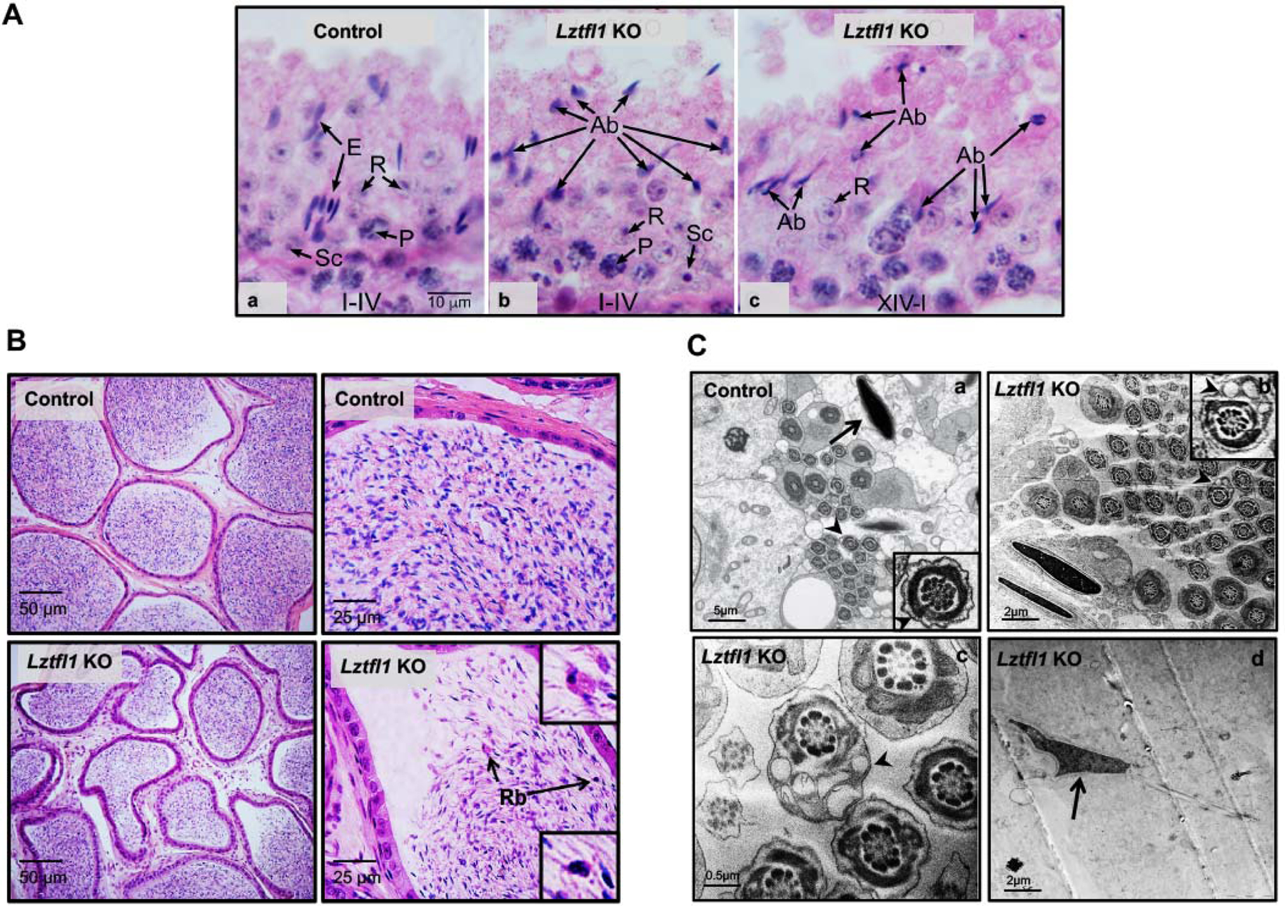
Figure 6. Histology and TEM analysis for the adult control (heterozygote) and Lztfl1 KO mice.

A. Testis from control and Lztfl1 KO mice. (a) In the control testis, stage I-IV tubule shows normal elongated spermatid heads (E) in bundles between the round spermatids (R). Some elongated heads are seen deep within the epithelium, between pachytene spermatocytes (P). Sc, Sertoli cell nucleus. Micron marker for all photos (10 μm). (b-c) Lztfl1 KO testis shows normal round spermatids (R) and pachytene spermatocytes (P), but the seminiferous epithelium contains numerous abnormal elongating spermatids (Ab). With H&E staining of stages XIV-IV, only abnormal elongating spermatid head shapes could be detected.
B. Numerous mature sperm were present in the cauda epididymal lumen of a control mouse (upper panel). But in the Lztfl1 KO mouse, there were fewer sperm in the epididymal lumen and residual bodies (Rb, arrows) appeared to be attached to numerous abnormal sperm (lower panel).
C. TEM images of elongated spermatids from control (a) and Lztfl1 KO (b-d) mice. In the control mice, nuclei have normal elongated shapes with condensed chromatin (arrow) and the flagella contains normal axonemes and fibrous sheath (arrow heads) within the seminiferous tubule. However, in Lztfl1 KO mice, large vacuoles were observed in the sperm flagella (arrow heads in b, c). The arrow in d point to abnormal shapes of nucleus in the Lztfl1 KO mouse.
