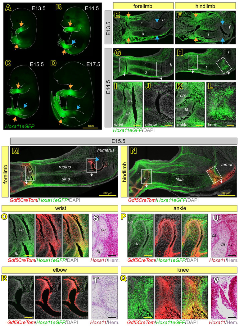
Fig 1. Hox11 is expressed throughout zeugopod embryonic joint development.
(A-D) Whole mount imaging of Hoxa11eGFP embryos at E13.5, E14.5, E15.5 and E17.5 show regional restriction to distal zeugopod elements of the limbs at all stages (A-D, orange arrows) and in the knee starting at E14.5 (B-D, blue arrows). (E-L) Tissue sections of Hoxa11eGFP embryos at E13.5 show Hox11 expression only in the wrist and ankle joints (E-F, orange arrows) and not in elbow or knee joints (E-F, blue arrows). At E14.5, expression in distal joints is maintained and Hox11 is additionally expressed in the knee (G-H). High magnification images from boxed areas in G and H show particularly high Hox11 expression in the scaphoid (sc) and lunate (lu) of the wrist (I) and talus (ta) and calcaneous (ca) of the ankle (K) and adjacent zeugopod long bones. Expression is also high in the E14.5 knee (L), but is largely absent in the elbow (J). (M-R) Tissue sections at E15.5 of the entire limb zeugopod show continuous Hox11 expression in the wrist (M), ankle and knee (N), but not elbow (M, blue arrow) and overlap with Gdf5Cre;tdTomato (Gdf5CreTom) joint progenitors (yellow). High magnification images of boxed regions in M and N show further restriction of Hoxa11eGFP to outer cells of the scaphoid (sc) and lunate (lu) in the wrist (O) and talus (ta) and calcaneous (ca) in the ankle (P) and also the high degree of overlap with Gdf5-lineage cells (Gdf5CreTom). Hoxa11eGFP also overlaps strongly with Gdf5CreTom in the knee (Q), but not in elbow (R). (S-V) RNAscope for Hoxa11 confirms expression patterns of Hoxa11eGFP in the wrist (S), elbow (T), ankle (U) and knee (V). r, radius; u, ulna; h, humerus; t, tibia; f, femur. scale bar = 100 μm (if not otherwise noted).

