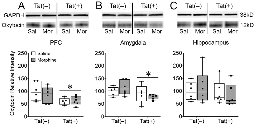
Fig. 4. HIV-1 Tat, but not morphine decreased PFC and amygdalar, but not hippocampal oxytocin levels.

After 8 weeks of Tat, 2 weeks of morphine exposure, and behavioral testing in assays of sociability Tat(+) mice, regardless of morphine exposure, exhibited lower expression of oxytocin in the PFC (A) and amygdala (B), but not the hippocampus (C) as measured by western blotting. Representative blots show decreased oxytocin in Tat(+) compared to Tat(−) mice in the PFC (A, top) and amygdala (B, top). All oxytocin western blots are represented as relative intensity to GAPDH normalized to Tat(−)/Saline. Data are presented as mean ± SEM; n = 9-10 mice per group. Main effect of Tat, *p < 0.05 vs Tat(−) mice.
