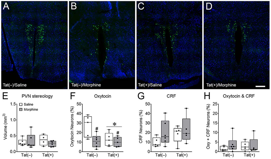
Fig. 6. HIV-1 Tat and morphine decrease hypothalamic paraventricular nucleus (PVN) oxytocin expression.

Representative images of cells immunoreactive for both oxytocin (green) and the neuronal marker NeuN (blue) in the PVN (A-D) imaged with Keyence VHX-7000 digital microscope at 40× magnification and stitched together. Mice exposed to Tat for 8 weeks, administered 2 weeks of morphine, and tested in social interaction assays did not demonstrate changes in hypothalamic PVN volume (E). Tat and morphine decreased oxytocin- (F), but not corticotropin releasing factor (CRF)- (G) or oxytocin- and CRF-immunoreactive colocalized (H) neurons in the PVN of the hypothalamus. These data suggest that oxytocin-expressing neurons in the PVN are selectively vulnerable to morphine and Tat, but do not show if this vulnerability is due to changes in the metabolism or release of oxytocin, or the loss of the oxytocin-expressing neuron subpopulation. Data are presented as mean ± SEM; n = 4-6 mice per group. Main effect of Tat, *p < 0.05 vs Tat(−) mice. Main effect of morphine, #p < 0.05 vs saline treated mice. Scale bar = 200 μm.
