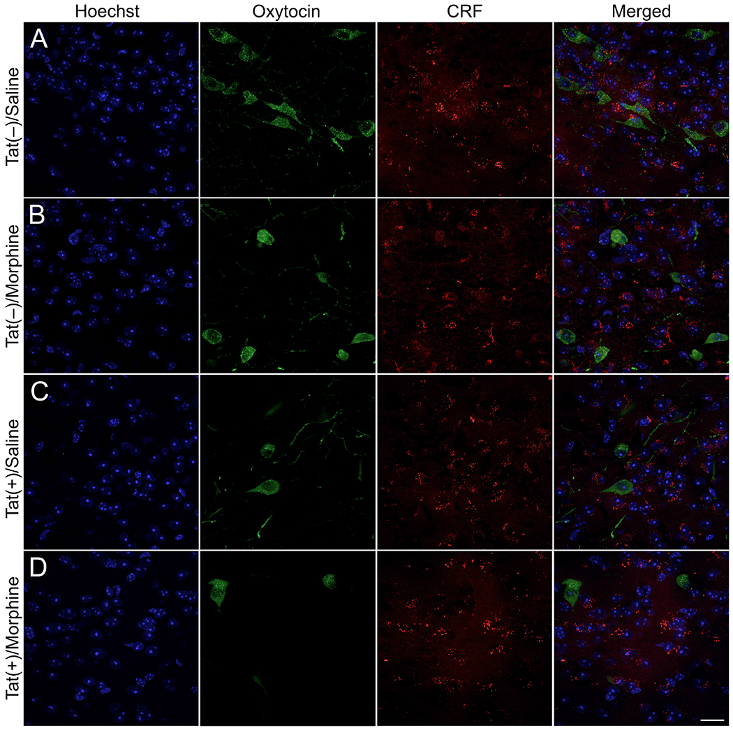
Fig. 7. Cellular localization of oxytocin, corticotropin releasing factor (CRF), and NeuN immunoreactivity in the hypothalamic paraventricular nucleus (PVN).

Representative images of oxytocin (green), CRF (red), and Hoechst (blue) positive cells were taken using a Zeiss LSM 700 microscope at 63× magnification (Zeiss, Oberkochen, Germany). Tat(−)/morphine (B), Tat(+)/saline (C), and Tat(+)/morphine (D) PVN tissue sections had less oxytocin immunoreactive-cells compared to Tat(−)/Saline (A) control sections. Scale bar = 20 μm.
