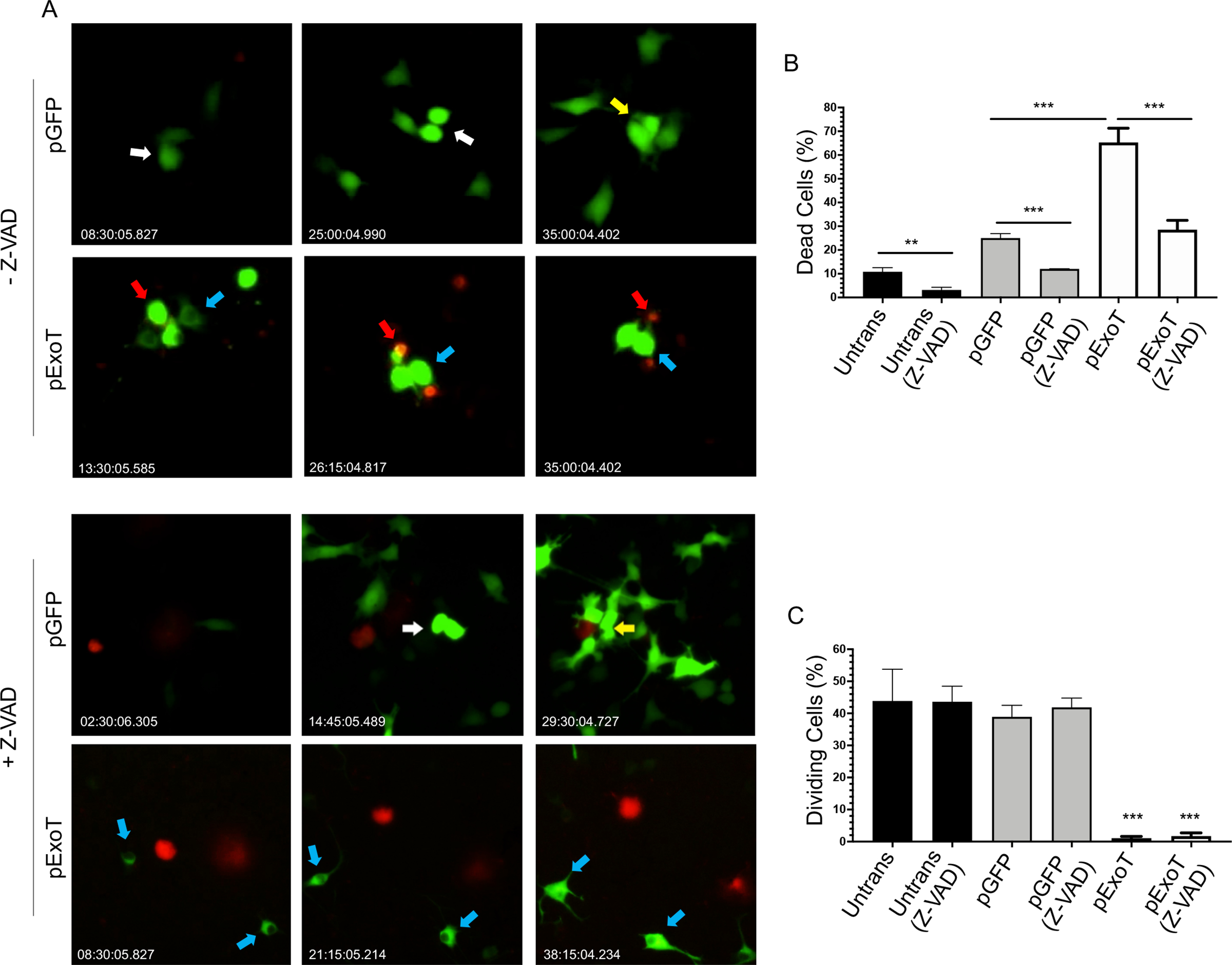
Fig. 1: Impact of ExoT on cell death and proliferation in B16 melanoma cells.

B16 cells were transfected with pIRES mammalian expression vector expressing either wild type ExoT, fused to GFP at the C-terminus (pExoT), or the vector control (pGFP), in the absence or presence of ZVAD (60μM) to block apoptosis. Cell death was analyzed by time-lapse video microscopy in the presence of the impermeant nuclear dye propidium iodide (PI) which stains dead cells (red or yellow). Proliferation was assessed by determining the percent of cells undergoing mitosis. A) Representative frames of videos are shown. Red arrows point to representative transfected cells that succumb to death. White arrows point to non-apoptotic transfected cells that undergo mitosis for the first time after transfection. Yellow arrows point to non-apoptotic transfected daughter cells that undergo mitosis for the second time after transfection. And blue arrows point to representative non-apoptotic transfected cells that fail to undergo mitosis. The tabulated results for cell death are shown as the Mean ± SEM in (B) and for proliferation (cell division) in (C). (N≥3, **p<0.01, ***p<0.001, Student’s t-test).
