
Fig. 3. Both domains of ExoT contribute to ExoT’s dampening of G1/S checkpoint regulators.
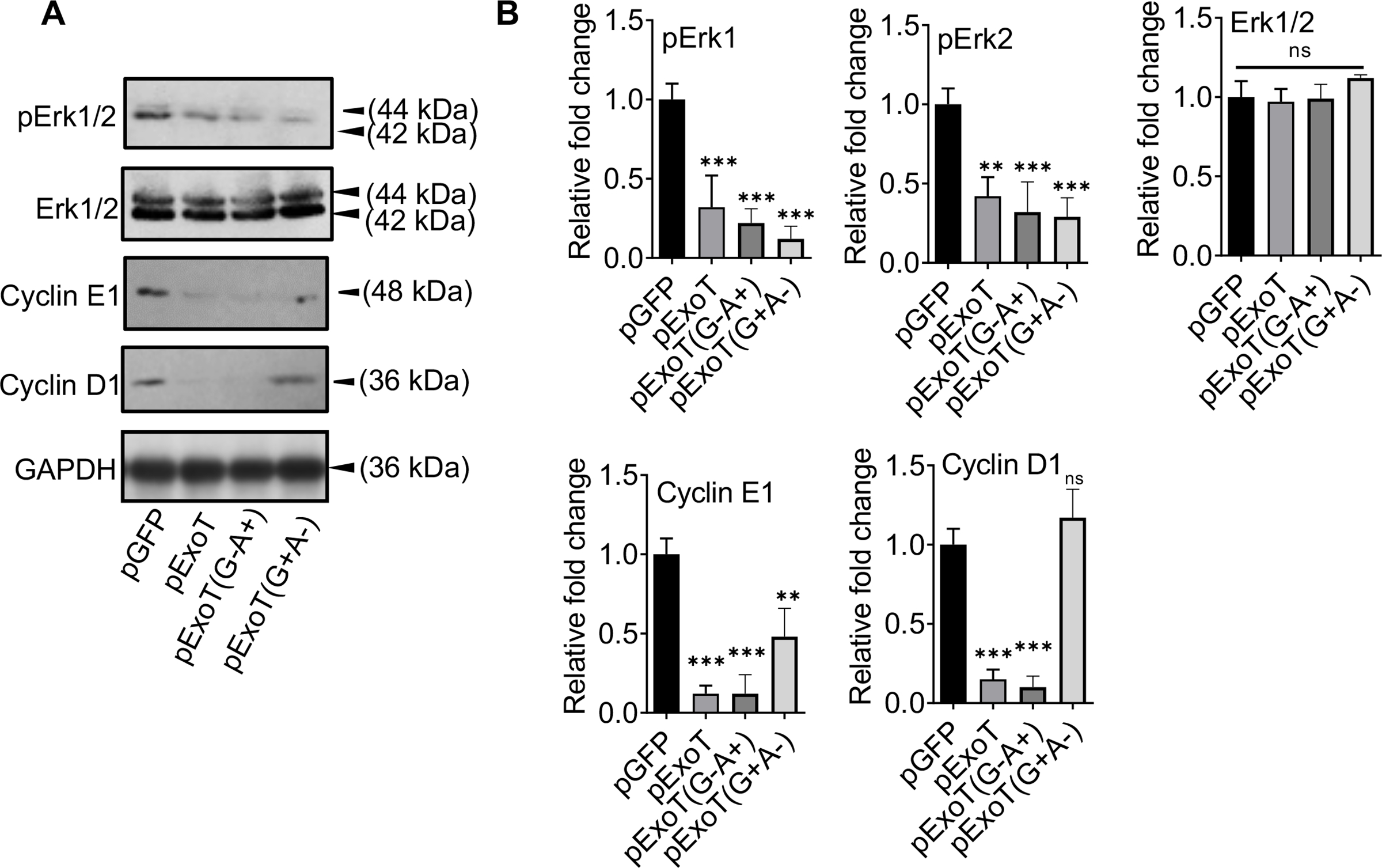
A) B16 cells were transfected with indicated expression vectors in the presence of Z-VAD. Cell lysates were assessed for the indicated G1/S checkpoint proteins, 48h after transfection by Western blotting. B) The corresponding densitometer data from 3 replicates, as compared to pGFP control vector, are shown after normalizing the data to their corresponding GAPDH levels. (N=3; ns, not significant, * p<0.05, ** p<0.01, *** p<0.001; One-way ANOVA).