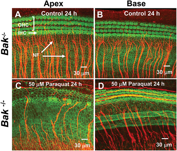
Figure 3:
Paraquat destroys hair cells and nerve fibers in the apical turn of Bak homozygous (Homo) mice. Representative confocal photomicrographs of cochlear organotypic cultures from the apex and base of the cochlea from a Bak−/− mouse double-labeled with phalloidin conjugated to Alexa 488 (green) to label the three parallel rows of outer hair cells (OHCs) and a single row of inner hair cells (IHCs) plus a primary antibody against neurofilament 200 conjugated to a secondary antibody (red) to label the nerve fibers (NFs) projecting out radially towards the hair cells. Note orderly rows of hair cells and large fascicles of nerve fibers in (A) apical turn and (B) basal turn of Control cochleae (0 μM paraquat) cultured for 24 h. After 24 h treatment with 50 μM of paraquat, nearly all OHCs, IHCs and NFs were missing in the (C) apex of the cochlea whereas (D) most hair cells and nerve fibers were present in the base of the cochlea.

