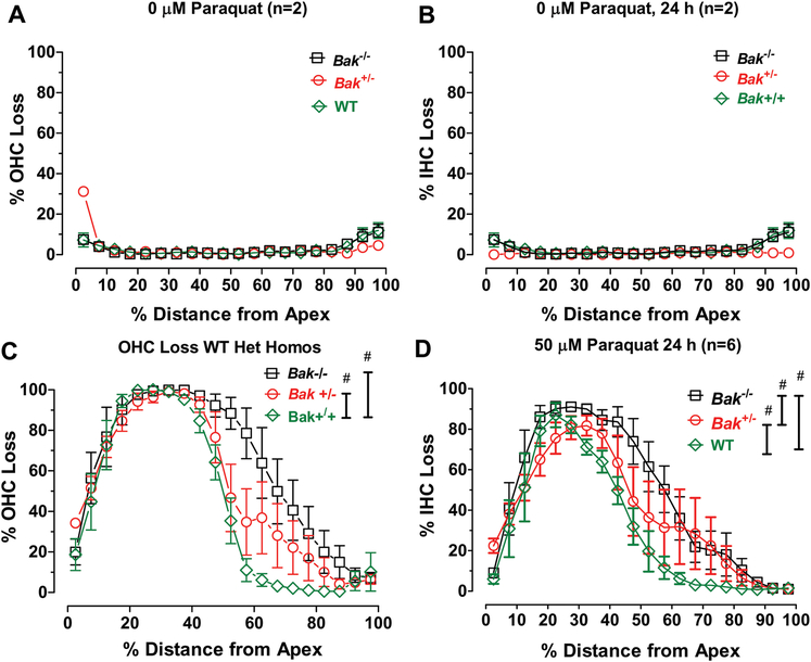
Figure 4:
Paraquat-induced hair cell lesions are greater in Bak heterozygous (Het) and homozygous (Homo) mice than Bak WT mice. Mean cochleograms showing percent OHC loss and percent IHC loss as a function of percent distance from apex of the cochlea of Bak WT, Het and Homo mice. Mean (+/− SEM) cochleograms from Control cochlea (0 μM paraquat) exhibit little OHC (A) or IHC (B) loss in Bak WT (n=2), Het (n=2) and Homo (n=2) mice. Mean (n=6/group, +/− SEM) cochleograms showing (C) OHC loss and (D) IHC loss in Bak WT, Het and Homo mice. # indicates significant difference (p<0.05) between groups designated by the vertical bars.

