Figure 7:
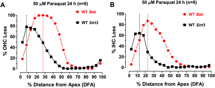
Bak WT mice are more vulnerable to paraquat ototoxicity than Sirt3 WT mice. (A) Mean OHC loss versus percent distance from the apex (DFA) in Bak WT mice compared to Sirt3 WT mice. Maximum OHC loss occurred 27.5% DFA in Bak mice versus. 7.5% DFA in Sirt3 mice. (B) Mean IHC loss versus percent DFA in Bak WT mice compared to Sirt3 WT mice. Maximum IHC loss occurred approximately 22.5% DFA in Bak mice versus. 12.5 DFA in Sirt3 mice. The width of the IHC lesion in Bak mice extended over much of the apical half of the cochlea; the peak loss, which approached 90%, occurred roughly 25% from the apex. The IHC lesion in the Sirt3 group was restricted to a narrower range in the apical 30% of the cochlea and the maximum loss approached 80% and occurred roughly 10% from the apex. Thus, the hair cells lesions in the Bak mice were broader, more severe and peaked slightly further from the apex than those in the Sirt3 mice.

