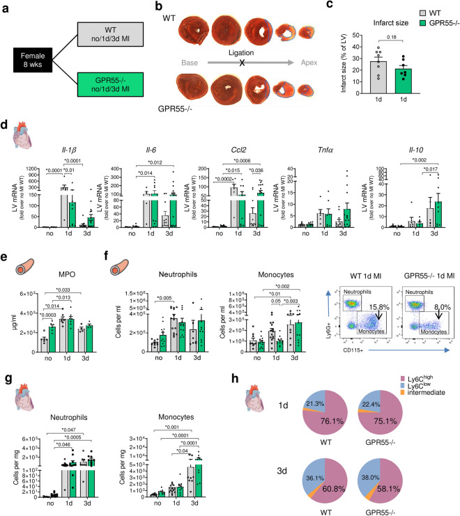
Figure 2.
Impact of GPR55 deficiency on acute inflammatory response following MI. (a) Study design to explore the effect of GPR55 deficiency on acute post-MI wound healing. WT and GPR55−/− mice were randomly assigned to baseline characterization (no MI) or to MI via permanent LAD ligation for 1 or 3 days (d). (b) Representative TTC stained consecutive heart slices from base to apex with dotted blue lines indicating the infarct area measured and (c) quantification of infarct sizes 1 day post-MI in WT and GPR55−/− hearts (student’s t-test). (d) LV gene expression in WT and GPR55−/− mice relative to baseline WT. (e) Plasma MPO levels in WT and GPR55−/− mice, assessed via ELISA. (f) Total counts of circulating neutrophils and monocytes in WT and GPR55−/− mice, quantified via flow cytometry, and representative dot plots indicating respective monocyte counts (CD115+ Ly6G−) as percentage of single cells. (g) Neutrophil and monocyte counts in WT and GPR55−/− hearts. (h) Relative distribution of Ly6Chigh, low and intermediate monocytes post-MI in WT and GPR55−/− mice. Bars indicate mean ± SE (n = 5–11/group) (Two-way ANOVA with Sidak’s post hoc test).

