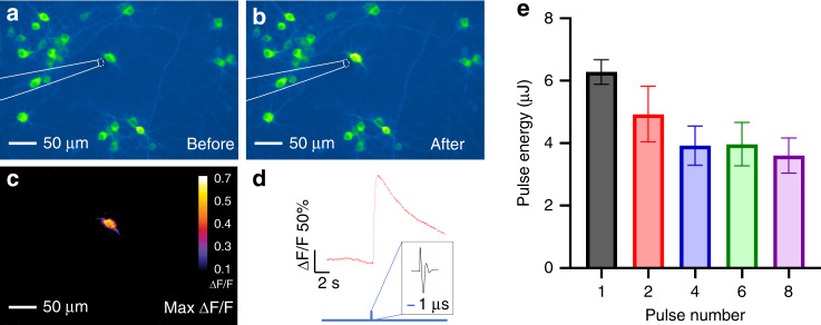
Fig. 3. Pulse energy dependence of TFOE stimulation.
a–c Fluorescence images of GCaMP6f expressing neurons before and after TFOE stimulation with a single pulse. d Calcium trace of the targeted neuron undergone single-pulse stimulation. Blue vertical line: onset of optoacoustic stimulation with zoom-in showing a representative optoacoustic waveform. e Pulse energy threshold for successful neuron stimulation as a function of pulse number (N = 5–7).

