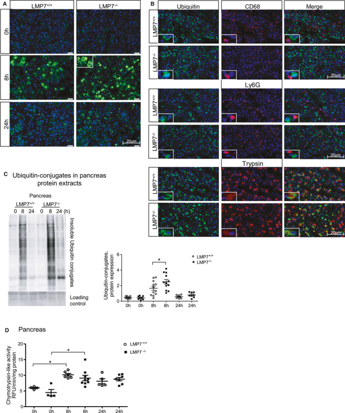
FIGURE 4.

Impairment of ubiquitinated protein degradation in pancreatic acini from LMP7−/− mice. Immunofluorescence in pancreas sections performed using ubiquitin A) alone or B) combined with CD68, Ly6G or trypsin antibodies. DAPI was used as nuclei staining. Scale bars, 20 µm. Image representative of five independent experiments. C, Higher accumulation of insoluble ubiquitin conjugates was quantified by Western blotting, and its ratio to a loading control (ponceau staining) was estimated by densitometry. (n = 8‐12). D, Proteasome‐like chymotrypsin activity was measured in pancreas homogenates using fluorescent substrate. Data are representative of independent experiments and expressed as means ± SEM. 0 h (n = 4), 8 h (n = 6‐9) and 24 h (n = 5‐7). *P < .05
