
FIGURE 2.
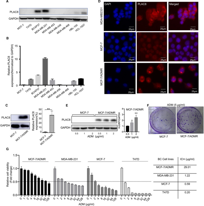
PLAC8 expression was increased in ADM resistance breast cancer cells. A, B. The protein and mRNA expression level of PLAC8 in various breast cancer cell lines. C Protein and mRNA expression of PLAC8 in MCF‐7 and MCF‐7/ADMR cells. D. Immunofluorescence staining for PLAC8 (red) and DAPI nuclear staining (blue) in MDA‐MB231, MCF‐7 and MCF‐7/ADMR cell lines. Scale bar = 25 µm. E. Altered PLAC8 expression in MCF‐7 and MCF‐7/ADMR cell upon ADM treatment (0.5, 1 and 2 µg/ml, 48 h). F. Colony formation indicated the proliferative potential of MCF‐7 and MCF‐7/ADMR cells with ADM treatment (5 µg/ml). G. The sensitivity of MCF‐7, MDA‐MB231, T47D and MCF‐7/ADMR cells to ADM. Each bar represents the mean ± SD of three independent experiments. *P < .05, **P < .01, ***P < .001