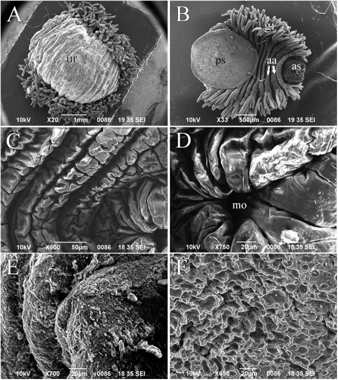
Fig. 5.

Scanning electron micrographs of O. jantseanus. (A) Entire body, dorsal view. Bulging on the back of the trachelosome (tr). (B) Entire body, ventral view. The posterior annulus was folded inside the anterior annulus (aa). (C) Trachelosome, ventral view. (D) Enlarged view of the mouth (mo). (E) Close-up of the dorsal view of O. jantseanus. (F) Close-up of the posterior sucker, ventral view. Scale bars: A, 1000 µm; B, 500 µm; C, 50 µm; D–F, 20 µm.
