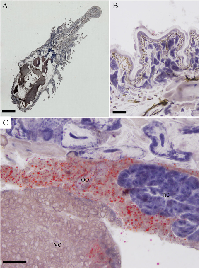
Fig. 7.

Sudan IV staining of frozen sections of O. jantseanus on the coronal plane. (A) General view of O. jantseanus. Substantial accumulation of lipid droplets was not observed. (B) Close-up view of the epidermal tissue. No lipid droplets were found. (C) Close-up view of the splanchnic tissue. Orange lipid droplets were found in the immature oocytes (oo), and a few lipid droplets were detected in vitelline cells (vc). No lipid droplets were detected in nurse cells (nc). Scale bars: A, 1000 µm; B-D, 50 µm.
