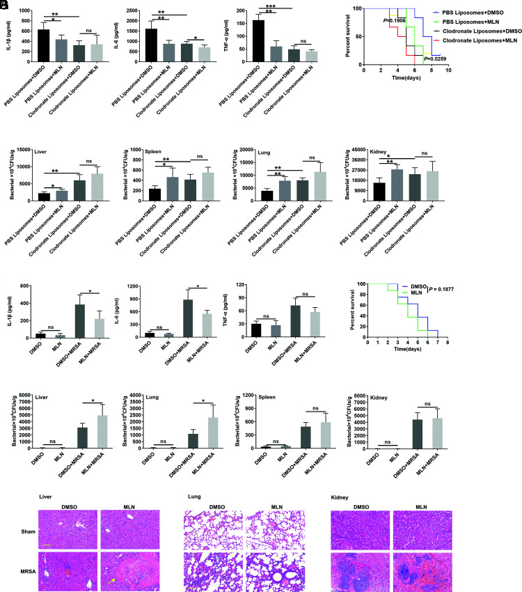
FIGURE 3.
Macrophages play an essential role in the effect of MLN4924. (A and B) Twelve hours after their i.v. injection of 50 μl of clodronate-encapsulated liposomes, mice were i.p. administered MLN4924 (30 mg/kg). Twelve hours after MLN4924 admission, 1 × 10⁸ CFUs of USA300 were injected into the mice through the tail vein. Lung, liver, kidney, and spleen tissues and blood were harvested 24 h later, and serum IL-1β, TNF-α, and IL-6 levels were measured by ELISA (A). The CFUs in the liver, lungs, spleen, and kidneys were measured (B) (n = 6). (C) Twelve hours after i.v. injection of 50 μl of clodronate-encapsulated liposomes, mice were i.p. administered MLN4924 (30 mg/kg). Twelve hours after MLN4924 admission, 5 × 10⁷ CFUs of USA300 were injected into the mice through the tail vein, and the 10-d survival rate was recorded (n = 6). (D–G) MLN4924 (100 nM)- or DMSO-treated BMDMs (4 × 10⁶ cells per mouse) were infused into the macrophage-depleted mice via tail veins. Twelve hours after macrophages infusion, USA300 (5 × 10⁷ CFUs per mouse for survival rate analysis, 1 × 10⁸ CFUs per mouse for others) was i.v. injected into the mice. Lung, liver, kidney, and spleen tissues and blood were harvested 24 h later, and serum IL-1β, TNF-α, and IL-6 levels were measured by ELISA (D). The CFUs in the liver, lungs, spleen, and kidneys were measured (E) (n = 5), inflammatory cell infiltration and abscess (yellow arrow) in the liver, lungs, and kidneys was visualized by H&E staining (F), and 8-d survival rate was recorded (G) (n = 8). Scale bars, 50 μm. Data are representative of three independent experiments (mean and SD). ns, not significant. *p < 0.05, **p < 0.01, ***p < 0.001 (unpaired Student t test).

