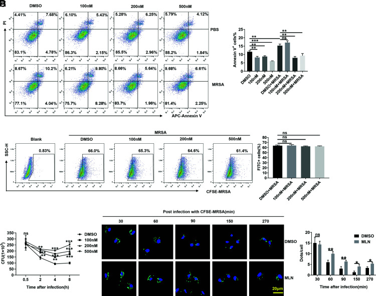
FIGURE 4.
MLN4924-treated macrophages are defective in USA300 killing. (A) PMs were treated with 100, 200, and 500 nM MLN4924 for 6 h, followed by infection with or without USA300 at an MOI of 20 for 6 h. Apoptotic cells were detected by flow cytometry (left) and statistically analyzed (right). (B) After treatment with 100, 200, and 500 nM MLN4924 for 6 h, PMs were infected with CFSE-labeled USA300 at an MOI of 20 for 30 min. CFSE-positive cells were analyzed by flow cytometry (left) and statistically analyzed (right). (C) After treatment with 100, 200, and 500 nM MLN4924 for 6 h, PMs were infected with CFSE-labeled USA300 at an MOI of 20 for 30 min. The infected cells were further cultured in sterile medium containing 2 µg/ml vancomycin, and CFUs were quantified at the indicated time points after USA300 infection. (D) PMs treated with MLN4924 (100 nM) or DMSO were infected with CFSE-USA300 at an MOI of 20 for 30 min and further cultured in sterile medium containing 2 µg/ml vancomycin. Then, the infected cells were washed, fixed, and stained with DAPI at the indicated times postinfection. CFSE-labeled USA300 (green) was visualized by fluorescence microscopy (left), and dots were analyzed (right). Scale bar, 20 μm. Data are representative of three independent experiments (mean and SD) (n = 3). ns, not significant. *p < 0.05, **p < 0.01, ***p < 0.001 (unpaired Student t test).

