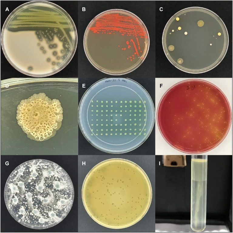
FIGURE 4.
Images captured with the imaging box. (A) Area of clearing on skim milk plates caused by protease secretion of P. aeruginosa. (B) Colonies of E. coli labeled with the red fluorescent protein DsRed-Express2. (C) Colonies of unknown microbes on an agar plate that was left open to the environment. (D) Close-up image of an unknown microbial colony. (E) Replica plate from the P. aeruginosa transposon mutant screening experiment. The three positions without a colony (B6, D8, and E7) represent non-growing mutant candidates. (F) A potato salad sample plated on mannitol salt plates to detect mannitol fermenting S. aureus, indicative as yellow colonies with yellow zones. (G) A plate of Penicillium sp. (H) Bacteriophage c2 plaques in a lawn of Lactococcus lactis. (I) Growth of Proteus vulgaris, a facultative anaerobe, in oxygen-reducing sodium thioglycolate medium, with dense growth in the high oxygen region and sparse growth in the low oxygen region.

