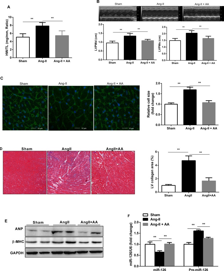
Fig. 2.
Asiatic Acid (AA) ameliorated AngII-induced cardiac hypertrophy and fibrosis in vivo. a Heart weight to tibia length ratio of different groups, n = 9. b Representative M-mode echocardiographic tracings of different groups. c Representative wheat germ agglutinin-stained of the left ventricles to cardiomyocyte size and quantification of the cardiomyocyte size in the indictated groups (n = 9 per group). d Representative Masson-staining of the left ventricles to assess cardiac fibrosis and quantification of the fibrosis area in different groups (n = 9 per group). e ANP (atrial natriuretic peptide) and B-MHC protein levels in the heart detected by Western blotting in different groups (n = 6 per group). f The expression of miRNA-126 and pre-miR-126 levels in the hearts of the different groups (n = 7 per group). Data are presented as the mean ± SD, **P < 0.01

