
Figure 1 |. VX2 tumor implantation and monitoring.
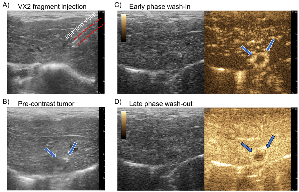
(A) An example B-mode image of the insertion of a 16-gauge coaxial biopsy needle into the liver parenchyma for VX2 tumor fragment injection. (B) After 2 weeks the implanted VX2 tumor (arrows) is difficult to visualize on B-mode imaging due to the similarity in echogenicity between liver and tumor. (C) During the early phase of contrast agent wash-in the VX2 tumor demonstrates a distinct rim enhancement on contrast-specific imaging. (D) The late-phase enhancement, representing portal vein blood flow, shows that the tumor has predominantly arteriole vascular supply.