Fig. 3.
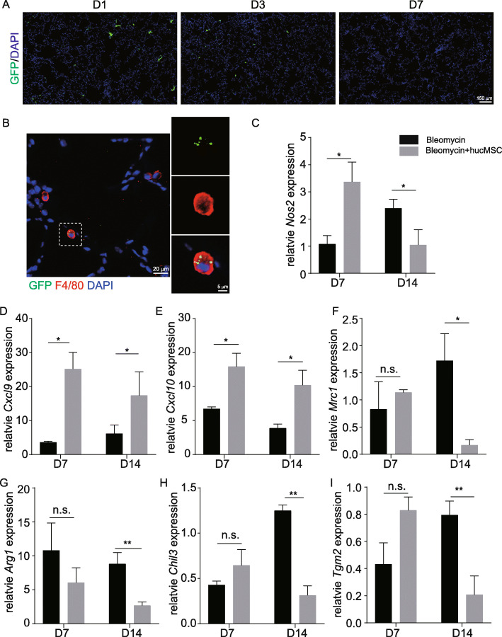
hucMSCs interacted with macrophages after infusion. A Immunofluorescent labeling of lung sections after GFP-labeled hucMSC infusion in bleomycin-treated lungs at various time points. B Lung sections were stained with antibodies against GFP and F4/80. C–I Nos2, Cxcl9, Cxcl10, Mrc1, Arg1, Chil3, and Tgm2 expression levels in bleomycin-treated lungs and hucMSC-treated lungs on day 7 and day 14 by whole lung qRT-PCR. Nos2, Cxcl9, and Cxcl10 expression levels (mean ± SD, n = 3 mice per group) were significantly increased in hucMSC-treated lungs compared to bleomycin-treated lungs at day 7 (C–E). Mrc1, Arg1, Chil3, and Tgm2 expression levels (mean ± SD, n = 3 mice per group) were significantly decreased in hucMSC-treated lungs compared to bleomycin-treated lungs at day 14 (F-I). The expression level of markers and cytokines in bleomycin-treated lungs and hucMSC-treated lungs was compared to saline-treated mice lungs. *p < 0.05, **p < 0.01, n.s. no significant difference, Student’s t-test

