
Figure 3.
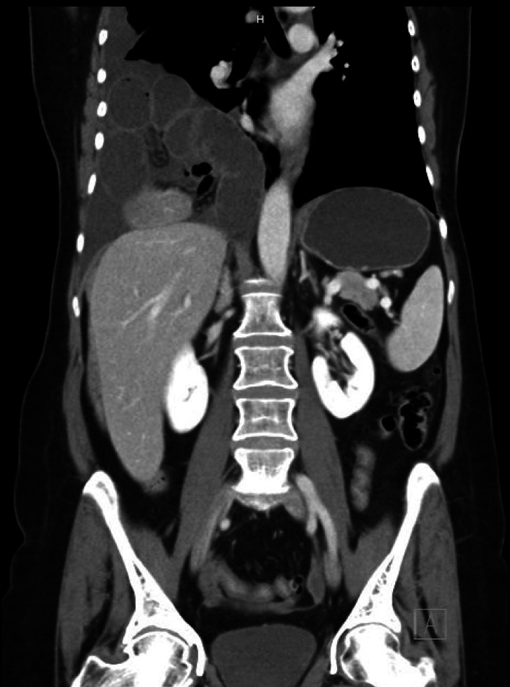
Contrast-Enhanced CT of the thorax, abdomen and pelvis in coronal view showed right anterior diaphragmatic defect (Morgagni hernia) with the evidence of dilated small bowel loops, mesenteric fat pad and part of the hepatic dome seen in right hemithorax. Also, subsequent appreciable dilatation of the stomach was noted.