FIGURE 1.
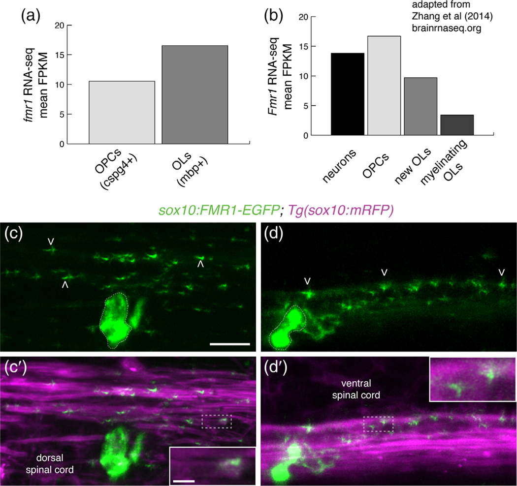
Fragile X mental retardation protein (FMRP) is localized within nascent myelin sheaths. (a) Zebrafish fmr1 expression levels (fragments per kilobase of transcript per million mapped reads; FPKM) from RNA-Seq of FAC-sorted cspg4+ olig2+ OPCs and mbpa+ olig2+ oligodendrocytes (Ravanelli et al., 2018). (b) Murine Fmr1 expression (FPKM) from RNA-Seq of neurons and oligodendrocyte lineage cells (Adapted from Zhang et al., 2014; brainseq.org). (c-c′) Lateral images of a living 4 dpf Tg(sox10:mRFP) transgenic larva transiently expressing sox10: FMR1-EGFP, a human FMR1-EGFP fusion construct, in an oligodendrocyte in the dorsal spinal cord. FMR1-EGFP expression is highest in the cell body (dashed outline), with dimmer, punctate expression noted in sox10:mRFP+ myelin sheaths of the dorsal spinal cord (arrowheads), including the terminal ends of sheaths (c′, inset). (d,d′) An oligodendrocyte in the ventral spinal cord transiently expressing FMR1-EGFP in a single myelin sheath on a large diameter Mauthner axon. The fusion protein is widespread in the cell soma (dashed outline) and punctate throughout the myelin sheath (arrowheads). FMR1-EGFP co-localizes with the sox10:mRFP+ myelin membrane (d′ inset). Wide scale bar = 10 μm, inset scale bar = 2 μm [Color figure can be viewed at wileyonlinelibrary.com]

