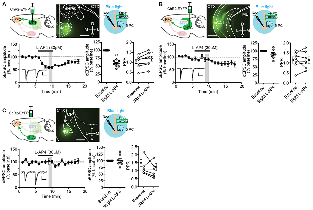
Fig. 1. Effects of L-AP4 on optogenetically evoked glutamatergic transmission at thalamo-, hippocampo-, and amygdala-mPFC synapses.

(A to C) Schematic diagram showing the injection of AAV5-CaMKIIa-hChR2(E123T/T159C)-EYFP into the mediodorsal thalamic nucleus (MDT; A), ventral hippocampus (vHPC; B), or basolateral amygdala (BLA; C), each with a representative image of EYFP-tagged ChR2 expression in the injection site 3 to 5 weeks after viral injection (calibration bar, 200μm), and a second schematic (right) depicting the acquisition of ex vivo whole-cell recordings from mPFC layer V pyramidal cells in response to light stimulation of ChR2:EYFP expressed exons. Bottom panels show the effect of L-AP4 on both oEPSCs over time and paired-pulse ratios (PPR) at each synapse type. Data are mean ± SEM from N = 8 neurons from total of 7 mice (A), and 7 neurons from 5 (B) or 4 (C) mice. Synapse-specific oEPSCs versus baselines were compared by Wilcoxon matched-pairs tests: **P <0.01 (A), P > 0.1 (B), and P > 0.9 (C). PPRs were also compared by Wilcoxon matched-pairs tests: **P < 0.01 (A), P > 0.9 (B), and P > 0.2 (C). Abbreviations: dHPC = dorsal hippocampus, CTX = cortex, vCA1= ventral CA1, MB = midbrain, CP = caudal putamen. Calibration bars for oEPSC traces: 100pA/20ms (A), 50pA/20ms (B), and 20pA/20ms (C). The gray-shaded vertical bar in the time course indicates the time points used to be averaged for statistical comparisons to baseline. For reference but not marked in the figure is the statistical comparison of oEPSCs and PPR at 14-15 min after L-AP4 application at hippocampo-mPFC synapses (B): P < 0.05 and P > 0.9, respectively, by Wilcoxon matched-pairs test.
