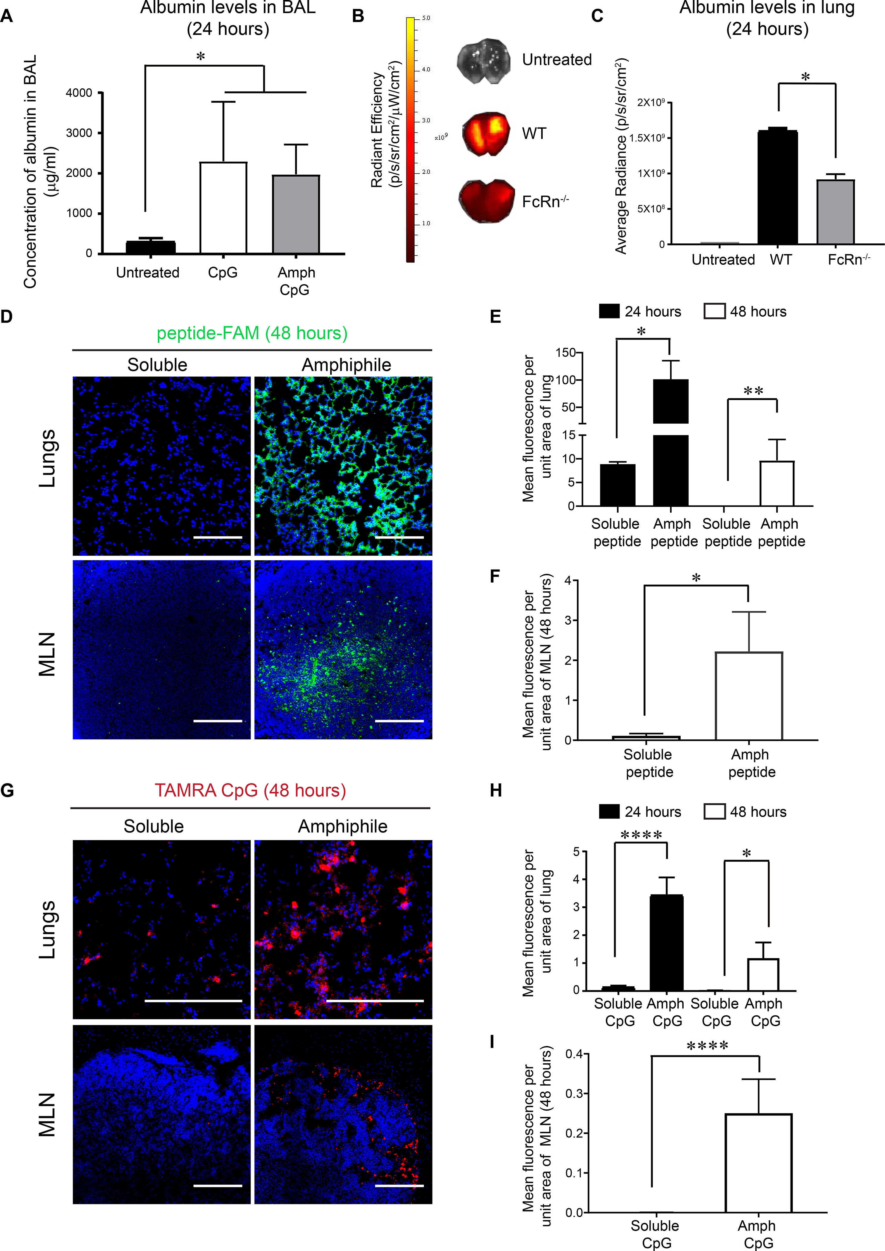
Figure 1. Amphiphile peptides or CpG are retained in the lung parenchyma and draining MLNs.

(A) Albumin levels measured by ELISA in bronchoalveolar lavage (BAL) from WT mice that were untreated or treated with inflammatory stimuli i.t. (B) Labeled mouse albumin was delivered i.t. into WT and FcRn−/− mice (n = 3/group) and fluorescence signal from lungs was measured by IVIS imaging 24 hours later. (C) Quantification of fluorescence signal from experiment described in (B). (D-I) C57BL/6 mice (n=3/group) were administered 20 μg FAM-labeled (green) gp100 peptide, an equimolar amount of labeled amph-gp100, 10 μg TAMRA-labeled (red) CpG, or an equimolar amount of labeled amph-CpG. Shown are representative immunofluorescence images of lungs (upper panel, scale bar = 100 μm in D and 200 μm in G) and MLNs (lower panel, scale bar = 100 μm) harvested from mice at 48 hours post immunization (D, G; DAPI (blue) was used to stain nuclei). Quantification of peptide or CpG signals from the lungs at 24 and 48 hours are shown in (E, H) and MLNs (F, I). Data shown are mean + SEM, *p<0.05, **p<0.001, ****p<0.0001 as determined by one-way ANOVA. Data are representative of at least two independent experiments.
