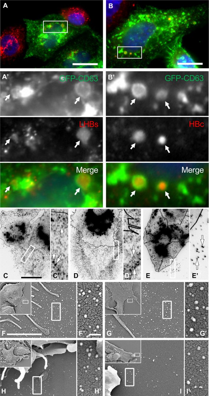
FIG. 1.

Association between intracellular CD63‐positive compartments and HBV proteins in HBV‐expressing cells that display CD63‐positive vesicular structures around cells. (A,A′,B,B′) HepG2.2.15 cells were transfected with GFP‐tagged CD63 (green) as a late endosome/MVB marker, then subsequently fixed and immunostained with antibodies to HBV components (red), including the LHBs protein (A′) or the HBc protein (B′). Arrows point to vesicles that are positive for both endosomal and viral antigens. DAPI (blue) for nuclear staining. (C‐E) Immunofluorescence images of HepG2.2.15 cells (C,D) or parental HepG2 cells (E) fixed and stained with antibodies to CD63. Images are presented as inverted gray scale to better contrast the substantial number of uniformly sized CD63‐positive vesicles surrounding each cell. Cells are outlined with black lines for clarity. Higher magnifications of boxed regions are provided in C′‐E′. (F,F′,G,G′,H,H′,I,I′) Scanning electron micrographs of cell borders showing the uniformity in size (40‐50 nm) of the HepG2.2.15 (F,F′,G,G′) and control HepG2 (H,H′,I,I′) generated vesicles. Scale bars: A‐C, 10 μm; F, 2 μm; F′, 200 nm.
