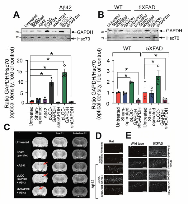
Figure 4.

GAPDH in the hippocampus promotes brain injury in AD animal models. (A) Representative Western blots of hippocampal samples from rats injected with vehicle (n = 3, sham-operated), lentiviral pLOC-GAPDH plasmid (n = 3), or lentiviral shGAPDH RNA (n = 3), with or without Aβ42. Hsc70 antibody was used as loading control (upper panel). GAPDH/Hsc70 ratio is shown as a fold change to control (untreated) for three animals in each experimental group (lower panel). (B) GAPDH western blots of hippocampal samples from WT and 5XFAD mice injected with vehicle (n = 3, sham-operated), lentiviral pLOC-GAPDH plasmid (n = 3), or lentiviral shGAPDH RNA (n = 3) (upper panel); quantitation of relative band intensity (lower panel). (C) Representative magnetic resonance brain images (of rats two months after injection of Aβ42 together with lentiviral pLOC-GAPDH or Aβ42 together with lentiviral shGAPDH RNA). MRI protocols: Rare T1, TurboRare T2, and FLASH (gradient echo (D) Representative frontal histological slices of hippocampus from experimental animals stained with the Click-IT TUNEL kit (n = 3 in each experimental group); Scale bar = 100 μm. (D) Representative histological slices (upper panel) of hippocampi of wt and 5XFAD mice, sham-operated or injected with lentiviral pLOC-GAPDH or shGAPDH RNA; Scale bar = 100 μm.
