
Figure 3.
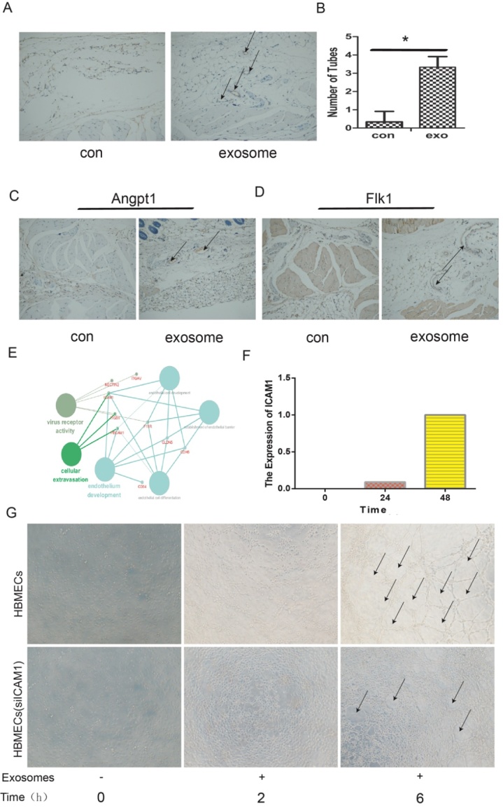
ICAM1 in HBMECs were upregulated after exosome treatment. (A) The number of tubes was detected by immune-histochemical staining (CD31). (B) The quantitative results (P<0.05). (C) Immunohistochemical staining results for the expression of ANGPT1 with control and exosomes stimulus. (D) The expression of FLK1 was detected using immunohistochemical with control and exosomes stimulus. (E-F) Proteomics and time series analysis of the target protein ICAM1, which was highly expressed after exosome stimulus. (G) The formation of tubes by HBMECs with or without ICAM1 knockdown.