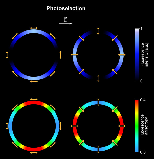
Figure 4.
Schematic representation of the idea of photoselection. Color rings represent the equatorial cross sections of the spherical lipid vesicles: imaged by means of fluorescence (top) or fluorescence anisotropy (bottom). Linear fluorophores represented by yellow arrows are bound to the membranes and oriented parallel (left) or perpendicular (right) with respect to the membrane plane of a lipid vesicle. The highest fluorescence signals and fluorescence anisotropy values are observed in the cases when the electric vector of the laser scanning light (E⃗) is parallel to the transition dipole of the light-absorbing molecules. According to this effect, linear chromophores oriented vertically to the membrane plane give rise to high fluorescence intensity and anisotropy levels on the left- and right-hand sides of the cross section of the lipid vesicle and the chromophores oriented horizontally to the membrane plane give rise to the high signals in the top and bottom parts of the liposome.

