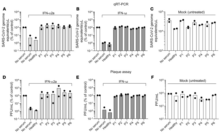
Figure 2. Auto-Abs in patients with APS-1 neutralize the ability of type I IFNs to inhibit SARS-CoV-2 infection.
Calu-3 cells were mock treated (no serum) or pretreated with indicated concentrations of human serum in the presence or absence of 200 IU/mL IFN-α2a (A and D) or 5 ng/mL IFN-ω (B and E) for 16 hours before infection. IFN and serum were removed, and cells were infected with SARS-CoV-2 at an MOI of 0.01 for 1 hour, washed, and fresh medium was applied to the cells. Twenty-four hours after infection, supernatant was harvested for viral RNA extraction and plaque assays. (A–C) Viral RNA was extracted from supernatant and SARS-CoV-2 genome equivalents/μL were quantified by qRT-PCR using primers targeting the E gene region. (D–F) Supernatants were titrated on Vero E6 cells and incubated for plaque formation for 3 days. Plaques were counted and PFU/mL was determined. Data were generated in 2 independent assays. Values obtained in the absence of serum and IFN were set to 100%.

