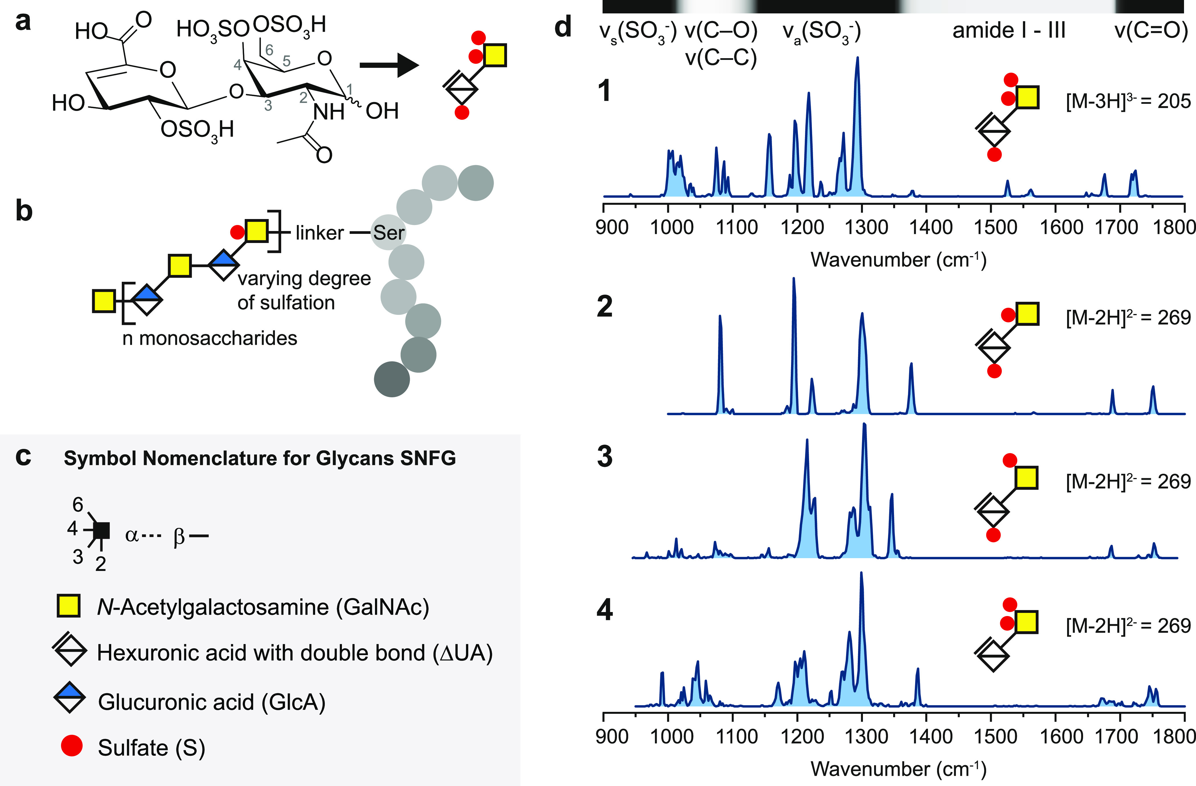
Figure 1.

(a) Investigated chondroitin sulfate disaccharide 1 in chemical representation followed by its representation in the symbol nomenclature for glycans (SNFG).37 Both α and β anomers are present in the sample. (b) Representative structure of the proteoglycan bikunin, which carries one site for an O-linked chondroitin sulfate chain.5,38−40 The protein chain is depicted with gray circles and the three-letter code is used to highlight serine. (c) SNFG. (d) Cryogenic IR spectra of triply sulfated disaccharide 1 investigated as a [M – 3H]3– anion with a mass-to-charge ratio (m/z) of 205 and doubly sulfated disaccharides 2–4 investigated as [M – 2H]2– isomeric anions with m/z of 269. On top of the first spectrum, the main vibrational features for these ions are qualitatively assigned in a horizontal bar.
