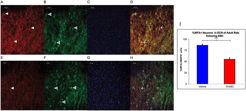
Fig. 2.
In Vivo Digestion of the PNN following EBC. A-D. Rats infused with vehicle show high WFA (red, panel A) reactivity with some PNN noted with white arrowheads as well as MAP2 reactive neurons (green, panel B) with some neurons noted with the open arrowheads and DAPI (blue, panel C) with all four channels merged in D. Three WFA+ neurons are marked with the winged arrowhead in D. E-H. Rats infused with ChABC have less WFA (red, panel E) labeling in comparison and in the merged image, there is only one WFA+ cell. I. A two-tailed unpaired t-test found ChABC infusion prior to EBC successfully decreased WFA reactivity in the adult rat AIN. *** = p < 0.001.

