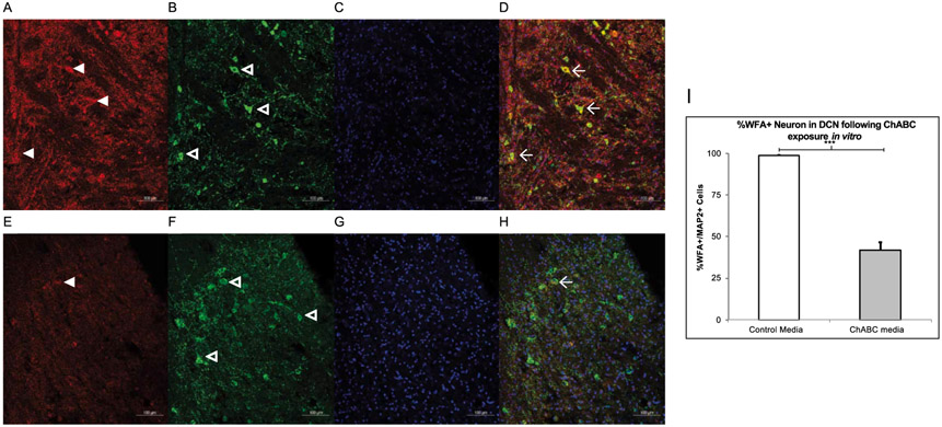
Fig. 9.
In Vitro Digestion of the PNN. A-D. Slices exposed with vehicle show high WFA (red, panel A) reactivity as well as MAP2 reactive neurons (green, panel B) and DAPI (blue, panel C) and all channels merged in D. Three WFA+ neurons are marked with the winged arrowhead in D. E-H. Slices exposed with ChABC have less WFA (red, panel E) labeling in comparison and in the merged image, there is only one WFA+ cell. I. A two-tailed paired t-test found ChABC exposure in the electrophysiological bath successfully decreased WFA reactivity in the juvenile rat AIN. *** = p < 0.001.

