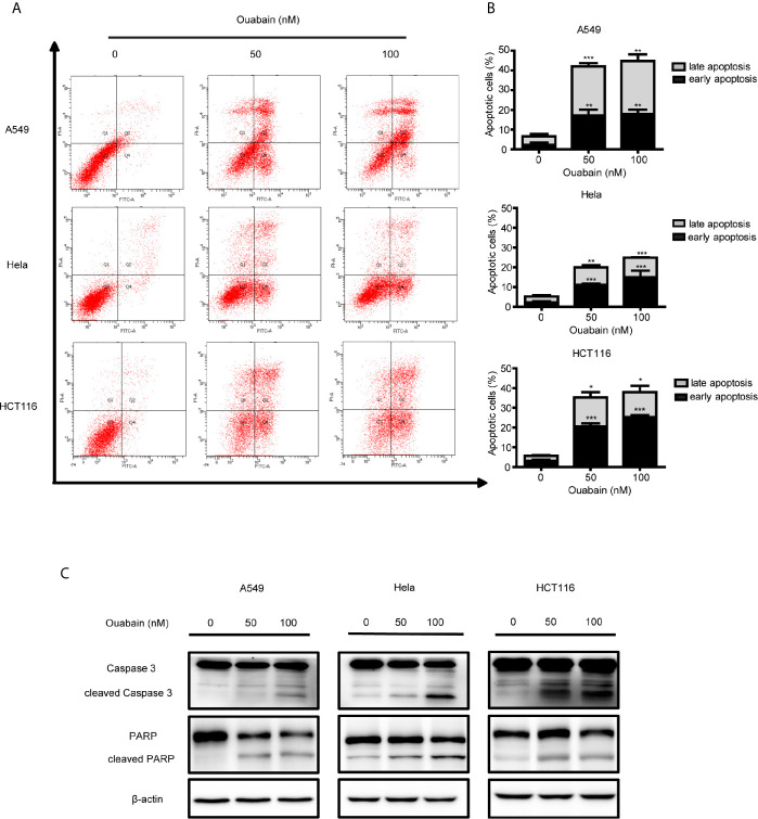
Figure 2.
Ouabain induced apoptosis of cancer cells. (A, B) A549, Hela and HCT116 cells were treated with indicated dose of ouabain (0, 50, 100 nM) for 48 hours, apoptosis was tested by flow cytometric analysis with Annexin V-FITC/PI staining. (C) The expressions of PARP, cl-PARP, Caspase3, cl-Caspase3 were analyzed by Western blotting. β-actin was used as a loading control. * P < 0.05; ** P < 0.01; *** P < 0.001 versus control group, n = 3.

