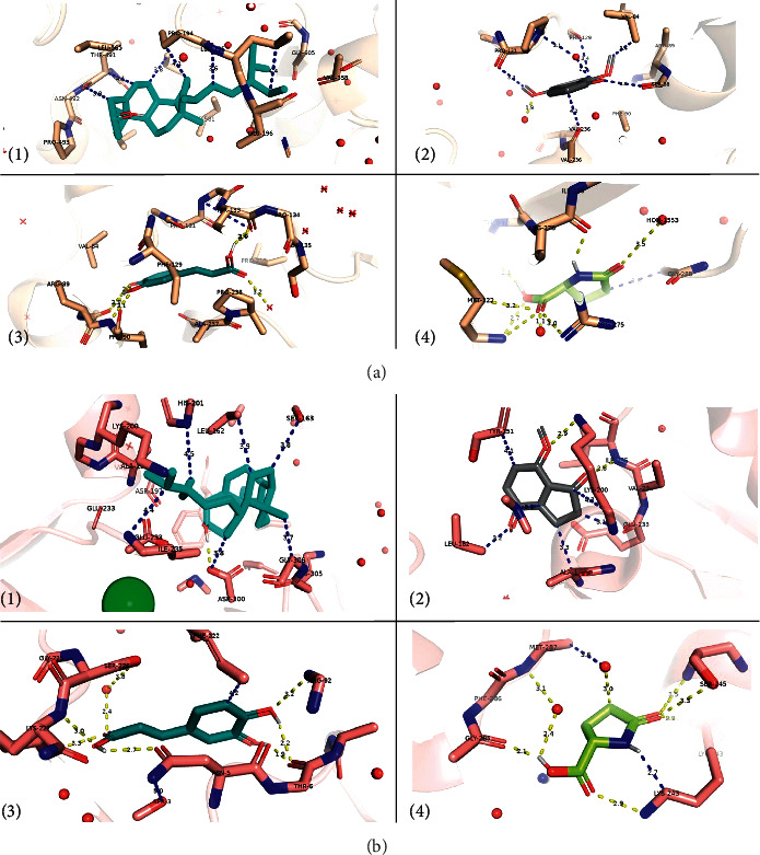
Figure 3.

Binding interface between plausible inhibitors and (a) the α-glucosidase enzymes: (1) beta-sitosterol, (2) 4,7-dimethoxyindan-1-one, (3) caffeic acid, and (4) pyroglutamic acid; and (b) the α-amylase enzymes: (1) beta-sitosterol, (2) 4,7-dimethoxyindan-1-one, (3) caffeic acid, and (4) pyroglutamic acid. All amino acids that are within 5 Ǻ from the ligand are shown as sticks. The rest of the protein is shown in an 80% transparent cartoon model. Polar contacts are shown in yellow, whereas other possible contacts are in blue. The green ball in label (1) of subpart 3(b) refers to a chloride ion near the active site.
