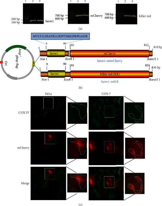
Figure 2.

Construction of mitochondrial targeting vector and localization. (a) PCR products of Sarm1, mCherry, and KR. Lane 1 was 100 bp DNA marker, and lanes 2 and 3 were PCR amplification products. (b) Schematic diagram of Sarm1-mtmCherry and Sarm1-mtKR vectors: MTS (Sarm1) was cloned into empty vector (plxsp-flag) (Not I and EcoR I sites), and mCherry and KR were cloned into plxsp-flag-Sarm1-MTS (EcoR I and BamH I sites). (c) The expressions of COX IV (green) and mCherry (red) were observed after COS-7 and HeLa cells were transfected with Sarm1-mtmCherry plasmids, ×200.
