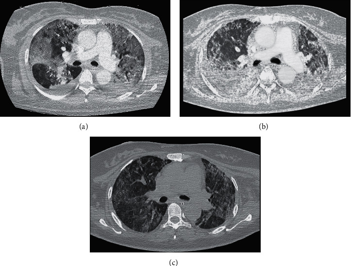
Figure 1.

Axial chest computed tomography scan. (a) Before the first extracorporeal membrane oxygenation support. Diffuse bilateral ground glass opacity pattern and right pleural effusion are shown. (b) During the first extracorporeal membrane oxygenation support. Thickening of interstitial septa and diffuse crazy paving are shown. (c) After the first decannulation of extracorporeal membrane oxygenation. Lung aeration improved bilaterally with partial resolution of ground glass opacities.
