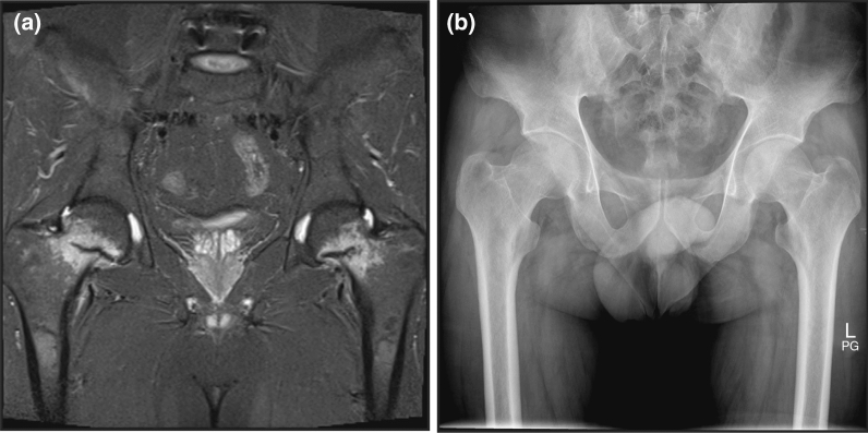
Fig. 2.
Patient diagnosed with symptomatic hypophosphatemia and osteomalacia with bilateral symmetric pseudofractures (looser zones) in the femur necks. a Magnetic resonance imaging showing marked hyperintensities of both femoral necks on T1-weighted imaging using a turbo inversion recovery magnitude sequence. Hyperintensities mark horizontal hypointensities extending halfway across the femoral neck. b X-ray plain film radiograph fails to show the fracture lines. Reprinted from Gastroenterology, 152(6), Benedikt Schaefer, Bernhard Glodny, Heinz Zoller, Blood and Bone Loser, e5–e6, Copyright (2017), with permission from Elsevier

