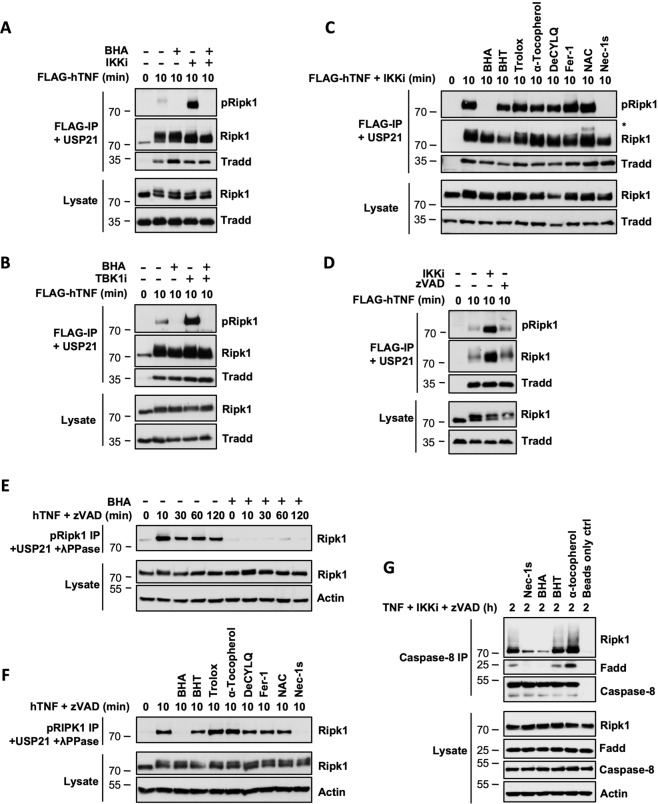
Fig. 3. BHA prevents cellular activation of RIPK1.
A–G MEFs were pretreated for 30 min with the indicated compounds (100 µM BHA, 100 µM BHT, 5 mM NAC, 10 µM DecylQ, 100 µM α-tocopherol, 100 µM Trolox, 500 nM Ferrostatin-1, 10 µM Nec-1s, 5 µM TPCA-1 (IKKi), 10 µM GSK8612 (TBK1i), 50 µM zVAD-fmk) before stimulation with 1 µg/ml FLAG-hTNF (A–D), 1 µg/ml hTNF (E–F) or 20 ng/ml hTNF (G) for the indicated duration. A–D TNFR1 complex I was FLAG-immunoprecipitated and the IPs were treated with USP21 before analysis by immunoblot. The signal for pRIPK1 refers to active RIPK1 autophosphorylated on residue S166/T169. E–F Autophosphorylated active RIPK1 (pRIPK1) was immunoprecipitated using the specific anti-pS166/T169 RIPK1 antibody and the IPs were treated with USP21 and λ phosphatase before analysis by immunoblot. G Complex IIb/necrosome was pulled down by immunoprecipitation of caspase-8 and analyzed by immunoblot. Immunoblots are representative of at least two independent experiments.

