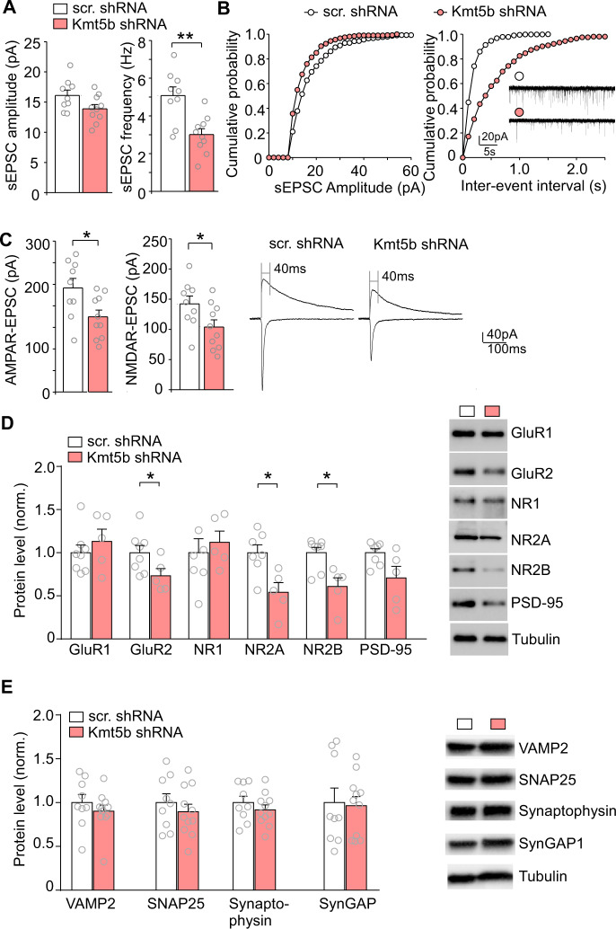
Fig. 3. Decreased synaptic transmission in Kmt5b-deficient PFC.
Bar graphs and cumulative probability plots of spontaneous EPSC (sEPSC) amplitude (A) and frequency (B) recorded from pyramidal neurons in the PFC of mice injected with Kmt5b shRNA or scr. shRNA AAV. Inset: representative sEPSC traces. C Bar graphs of evoked AMPAR- and NMDAR-EPSC amplitudes recorded from pyramidal neurons in the PFC of mice injected with Kmt5b shRNA or scr. shRNA AAV. Inset: representative eEPSC traces. D, E Bar graphs showing the expression level of synaptic proteins (normalized to tubulin) in the PFC from mice injected with Kmt5b shRNA or scr. shRNA AAV. Inset: representative Western blotting images. In all figures, *p < 0.05, **p < 0.01, t-test.

