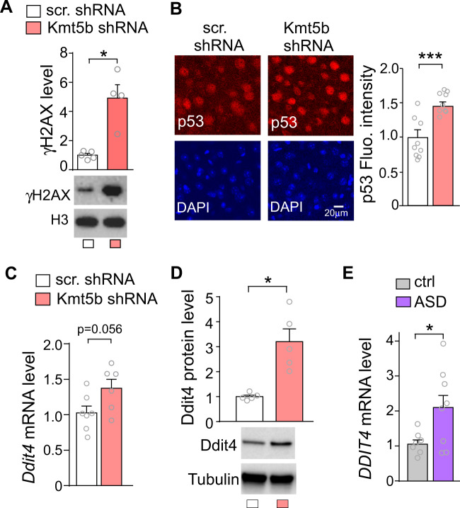
Fig. 4. Increased DNA damage, p53, and Ddit4 expression in Kmt5b-deficient PFC.
A Bar graphs and representative Western blots of γH2AX in the PFC of mice injected with Kmt5b shRNA or scr. shRNA AAV. B Immunocytochemical images and quantification of p53 in the PFC of mice injected with Kmt5b shRNA or scr. shRNA AAV. Bar graphs of Ddit4 mRNA (C) and protein (D) in the PFC of mice injected with Kmt5b shRNA or scr. shRNA AAV. E Bar graphs of DDIT4 mRNA in the PFC of postmortem human control vs. ASD patients. In all figures, *p < 0.05, ***p < 0.001, t-test.

