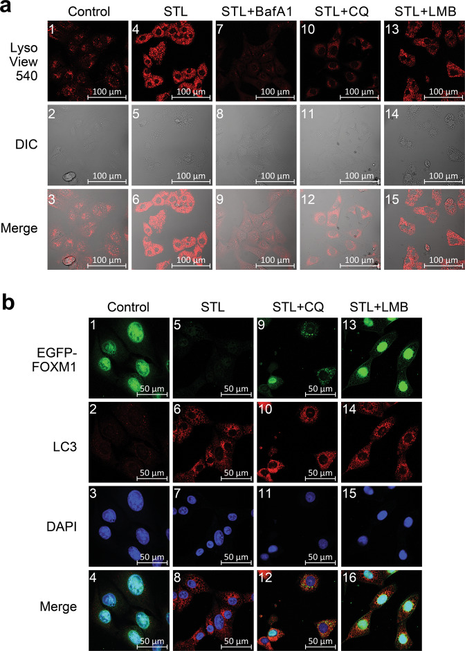
Fig. 4. STL promotes active lysosome formation and FOXM1 translocation from the nucleus to the cytoplasmic autophagosomes.
a Doxycycline-stimulated C3-luc cells expressing EGFP-FOXM1 fusion protein were treated with vehicle (“Control”, panels 1–3), 50 μM STL alone (“STL”, panels 4–6), or 50 μM STL in combinations with 25 nM BafA1 (“STL + BafA1”, panels 7–9), 40 μM CQ (“STL + CQ”, panels 10–12), and 25 nM LMB (“STL + LMB”, panels 13–15) for 12 h. Lysosomes were stained with vital LysoView 540 dye (red), cell morphology was analyzed using differential interference contrast (DIC) microscopy. b Doxycycline-stimulated C3-luc cells were treated with vehicle (“Control”, panels 1–4), 50 μM STL alone (“STL”, panels 5–8), or 50 μM STL in combinations with 40 μM CQ (“STL + CQ”, panels 9–12) and 25 nM LMB (“STL + LMB”, panels 13–16) for 24 h. Cells were stained for LC3 protein, nuclei were counterstained with DAPI. EGFP-FOXM1 (green), LC3 (red) and DAPI (blue) fluorescence was analyzed using confocal laser-scanning microscopy.

