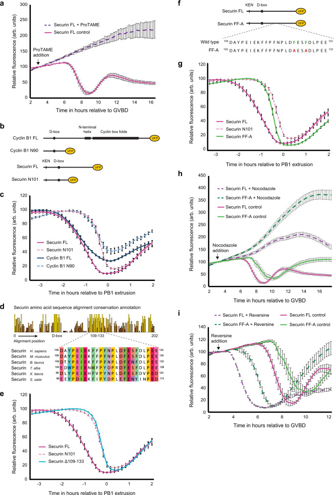
Fig. 2. A discrete region within the C-terminus of securin promotes destruction in prometaphase.
a Mean VFP-tagged securin FL (purple dashed trace, n = 11) destruction profile following incubation in 1.5 µM ProTAME to inhibit APC/C activity. Mean VFP-tagged securin FL (magenta trace, n = 16) destruction profile in control oocytes is included as a reference. ProTAME treated oocytes do not extrude a polar body and are therefore aligned at GVBD. b Schematic showing VFP-tagged securin and cyclin B1 constructs. c Mean securin FL (magenta trace, n = 25), securin N101 (pink dashed trace, n = 23), cyclin B1 FL (blue trace, n = 33) and cyclin B1 N90 (light blue dashed trace, n = 34) destruction profiles relative to PB1 extrusion. d Full sequence alignment conservation annotation and multiple sequence alignment of residues 109–133 in securin orthologs. e Mean VFP-tagged securin FL (magenta trace, n = 25), securin N101 (pink dashed trace, n = 23) and securin Δ109–133 (light blue trace, n = 23) destruction profiles relative to PB1 extrusion. f Schematic showing the position of Securin FF-A amino-acid substitutions. Residues F125 and F128 (shown in green in the wild-type protein) were switched to alanines (shown in red in Securin FF-A). g Mean VFP-tagged securin FL (magenta trace, n = 25), securin N101 (pink dashed trace, n = 23) and securin FF-A (green trace, n = 20) destruction traces relative to PB1 extrusion. h Mean VFP-tagged securin FL (magenta dashed trace, n = 23) and securin FF-A (dark green dashed trace, n = 30) destruction profiles following incubation in 150 nM nocodazole to arrest oocytes in prometaphase. Mean VFP-tagged securin FL (magenta trace, n = 16) and securin FF-A (green trace, n = 16) destruction profiles in control oocytes are included as a reference. Nocodazole treated oocytes do not extrude a polar body and are therefore aligned at GVBD. i Mean VFP-tagged securin FL (purple dashed trace, n = 16) and securin FF-A (dark green dashed trace, n = 16) destruction profiles following incubation in 100 nM reversine to block assembly of new SAC complexes. Mean VFP-tagged securin FL (magenta trace, n = 16) and securin FF-A (green trace, n = 16) destruction profiles in control oocytes are included as a reference. Reversine treated oocytes have an accelerated progression through meiosis and are therefore aligned at GVBD. All n numbers refer to the number of individual oocytes analysed over a minimum of three independent experiments. All error bars ± SEM.

